10 Revealing Scans of Historical Artifacts

In the not-too-distant past, when people were curious about the insides of artifacts or remains they just opened them up and went to town. Mummies were unwrapped and dissected, corroded containers pried apart, and the contents of vessels fished out with little regard for long-term preservation. Archaeologists and conservators are far more cautious today, and technology has made non-invasive examination possible in such detail that using your eyeballs pales in comparison. Here are 10 amazing views of history's innards.
1. MERESAMUN
CT scan of Meresamun wrapped in linen bandages. Image credit: The Oriental Institute via The History Blog.
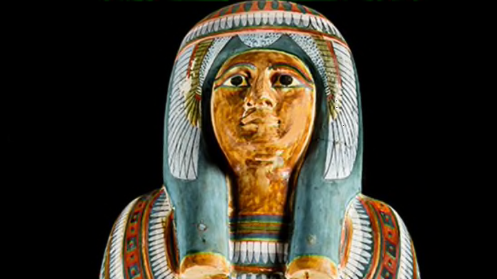
Meresamun was a singer in the Precinct of Amun-Re in Thebes's Karnak Temple Complex and was about 30 years old when she died in 800 BCE. Her mummy, sealed inside a richly painted cartonnage coffin (pictured at top), has been at the University of Chicago's Oriental Institute since 1920. Thankfully nobody tried to open the coffin, because it was so tightly sealed the beautiful exterior would have been damaged.
In 2009, Meresamun was given a CT scan at the University of Chicago Medical Center using a 256-slice scanner, which made it possible to virtually unwrap every layer, from the coffin down through the linen bandages, the surviving skin, tendons, and muscles, the packing materials embalmers placed inside her body, to the skeleton. They discovered she had all of her teeth, including her wisdom teeth, and not a single cavity. She also had a bunion on her right big toe. Whatever killed her, it doesn't show on her remains. You can see some of the thousands of Meresamun's CT scanner images on the Oriental Institute's website.
2. SOBEK
Trustees of the British Museum via The History Blog
The British Museum has a mummified Nile crocodile that was discovered at the temple at Kom Ombo, where the animals were raised and tended with care as the living incarnations of the god Sobek. Dating to between 650 and 550 BCE, the crocodile is huge, more than 13 feet long, and has 25 mummified baby crocodiles attached to his back, symbols of the deity's attributes of fertility and protection. For an exhibition that closed earlier this year, the British Museum had their Sobek CT-scanned, but it's not just any hospital that has the facilities to accommodate so large a patient. Hardened with a thick coating of resin, the mummy can't bend, so even carrying it past corners is a challenge; never mind finding a machine that could fit a giant Nile crocodile.
Fortunately, the Royal Veterinary College London's Equine Hospital had corridors and a CT scanner just big enough to do the trick. Their scans found that unlike most mummies, this croc was embalmed with his digestive organs intact, complete with the remains of his last meal. He ate well, too. There's a cow's shoulder bone and parts of a forelimb in his stomach, plus stones he'd eaten for ballast and to aid in digestion. The high-resolution scans were used to make 3D models of the layers of the crocodile's insides, which were put on display alongside the mummy in an exhibition called Scanning Sobek.
3. YOUNGEST-KNOWN MUMMIFIED FETUS
The Fitzwilliam Museum via The History Blog
The University of Cambridge's Fitzwilliam Museum recently scanned the contents of a miniature Late Period Egyptian coffin in their collection in preparation for the museum's Death on the Nile: Uncovering the Afterlife of Ancient Egypt exhibition. The finely carved cedar wood coffin dates to between 664 and 525 BCE and has been in the Fitzwilliam Museum since it was excavated in Giza in 1907. Inside is a bundle of linen bandages coated in black resin. The package was so small—the coffin is just 17 inches long—that curators thought it must have contained embalmed organs.
Before putting the coffin on display in the exhibition, curators decided to have a look at what was inside the bundle. An X-ray proved inconclusive, but a micro CT scan revealed the remains of the youngest-known mummified fetus. The skull and pelvis had collapsed, but all the fingers and toes were visible on the scan, as were the long bones of the arms and legs. Based on the bone length, radiologists were able to determine that the fetus was 16 to 18 weeks gestation. Few mummified fetuses have been found in Egypt, and this is by far the youngest. The two fetuses found in the tomb of King Tutankhamun were about 25 weeks and 37 weeks gestation.
The CT shows the little one's arms crossed over the chest, like pharaohs' arms were during the New Kingdom. The quality of mummification in the Late Period took a steep dive, with many mummies missing parts, or being a jumble of disarticulated bones shaped like a body. But this fetus was sent to the afterworld with the best care possible.
4. BRAIN REMOVAL STICK LEFT IN CRANIUM
RadioGraphics via The History Blog
When the Archaeological Museum in Zagreb, Croatia took a mummy they'd had since the 19th century to Zagreb's University Hospital Dubrava for a scan, they just wanted to find out more about it. CT scans, X-rays, and radiocarbon dating identified it as the mummy of a woman about 40 years old who died about 2400 years ago. But the CT scan also found something else: a tubular object stuck in her cranium. The researchers couldn't tell what it was made out of from the scan, so, with CT monitoring showing them the way, they sent an endoscope through the nasal cavity for further inspection.
The hole through the nasal cavity was made by the ancient embalmers so they could remove the brain. As it happens, it seems that this particular time they left the tool they'd used to liquify or remove the brain bits inside the cranium. The tool is made of cane or bamboo and it is one of only two brain removal tools ever discovered inside a mummy.
5. PLASTER CASTS OF VESUVIUS VICTIMS
CT scan of child's cast. Image courtesy the Archeological Site of Pompeii via the History Blog.
Vesuvius erupted on August 24, 79 CE, raining fine ash and pumice on the panicked population of Pompeii and then following it up with a deadly pyroclastic flow that sealed the ash and pumice layer, as well as any humans caught beneath it. The ash, lava, and gasses of the pyroclastic flow quickly hardened around the bodies. Over time, the soft tissues of the bodies decayed, leaving imprints in the ash layers alongside skeletons.
Centuries later, when excavations began, workers began noticing cavities with these imprints. In February 1863, pioneering archaeologist Giuseppe Fiorelli had the idea to fill these cavities with plaster and chip away at the hardened volcanic ash, leaving eerily detailed casts of Vesuvius' victims captured in the moment of their final agony.
Some of the plaster casts were recently restored and CT-scanned for the first time. The scans found that Pompeiians (at least the small sample cast in plaster and restored) had excellent teeth with nary a single cavity among them, in part due to the naturally high fluoride content of the water. The scans also revealed details about the remains and artifacts that had never been seen before because they were encased in plaster.
Here is the cast of a young child found next to a mother holding her baby in the House of the Golden Bracelet. The scan found that his full skeleton was intact in the plaster, that he was two to three years old, and that a bump on his sternum previously believed to be a knot in his clothing is in fact a gold fibula.
6. ROMAN CHAIN MAIL
X-ray of Harzhorn chain mail fragment. Image credit: Detlef Bach, Winterbach via The History Blog
For many years historians only knew what the long chain mail shirt worn by Roman soldiers, the lorica hamata, looked like from artistic depictions, such as the reliefs on the Ludovisi Battle Sarcophagus and Trajan's Column. The details of construction were elusive until archaeological remains of Roman chain mail were discovered starting in the 19th century.
Some finds are unmistakable, such as the mail shirts found in the barracks of the Roman fort of Arbeia in England, which are in exceptional condition. More frequently, chain mail finds are small fragments of corroded iron obscured inside a clump of soil. The pieces found at a 3rd century battlefield in Harzhorn, Germany, were so corroded that it was hard to tell with the naked eye that they were chain mail at all, except for the rippling pattern in the dirt encasing them. So archaeologists looked inside, where the x-rays revealed the small loops and intricate structure of the chain mail.
7. VIKING HOARD IN A CAROLINGIAN POT
Historical Environment Scotland via The History Blog
When archaeologists excavated the trove of Viking silver discovered by metal detector enthusiasts in Dumfries and Galloway, Scotland, in 2014, they unearthed more than 100 precious objects—silver ingots, solid gold and silver jewelry, glass beads—from multiple countries and cultures. Buried deep in a second excavated layer was a silver alloy pot with its lid still in place and sealed shut. The form and decoration of the vessel identified it as a piece of Carolingian manufacture made in western Europe between 780 and 900 CE.
The pot had turned green from the corrosion of copper in the silver alloy, and archaeologists didn't want to risk opening the lid and digging around inside without first having a clear idea of what was in there. Enter the Borders General Hospital and its CT scanner. The scans revealed an Anglo-Saxon openwork brooch, four more silver brooches, gold ingots, and ivory beads coated in gold, each piece individually wrapped in a protective textile or other organic material.
8. MUMMY INSIDE BUDDHA STATUE
The Drents Museum via The History Blog
A statue of the meditating Buddha with a mummy snugly fitted inside was scanned in 2014, before its appearance at the traveling Mummies of the World exhibition, then at the Drents Museum in the Netherlands. The statue is from China, and it's the only Chinese Buddhist mummy to be made available for scientific study in the West. While researchers knew that the statue contained a mummy, the CT scan taken at the Meander Medical Center in Amersfoort, central Netherlands, revealed unexpected contents nonetheless.
This was believed to be the mummy of Master Liuquan of the Chinese Meditation School, who died around 1100 CE after subjecting his body to an inconceivably grueling six-year process of starvation, dehydration, and poisoning with the sap used to make lacquer. The idea was to mummify the body while still alive until at the end the self-mummifier was walled in to a tight enclosure with enough air to breathe until he died. Three years later, if the body was found to be truly mummified, the monk was considered to have achieved enlightenment and was elevated to the rank of Buddha.
The CT scan found paper with Chinese characters written on it stuffed into the mummy where the internal organs would have been. Obviously, that cannot happen in the usual self-mummification process, which means this Buddha was at least in part mummified by embalmers, just like Meresamun and Sobek were.
9. A LITTLE BOX MEETS A SUPERPOWERED X-RAY
found next to a body buried in the crypt of the former Saint-Laurent church in Grenoble, France, is so corroded conservators wouldn't even attempt opening it to identify its contents. It's a wee cylindrical piece just an inch and a half in diameter that dates to the 17th century. Corrosion had eaten a hole in the lid, so conservators knew there were three circular objects inside. They thought perhaps they were coins, but it was impossible to make out details.
Even though it's a modest piece of no momentous historical import, conservators at the Grenoble Archaeological Museum asked their neighbors at the European Synchrotron Radiation Facility to use their synchrotron-generated X-rays which produce a light 100 billion times brighter than hospital X-rays to scan the little box. It was just a test, really, a feasibility study that would give the museum a cool picture for an upcoming exhibit. The results were so astonishing it turned into a full-blown research project.
The three circular objects were stuck together and in terrible condition, but the synchrotron scan was able to virtually peel them apart and show conservators every detail in extreme closeup. They're clay religious medallions, not coins, with images of Christ, the Virgin Mary, and the Nativity, and Latin inscriptions from the Bible and Catholic liturgy.
10. SPACESUITS
The National Air and Space Museum via The History Blog
In 2010, the Smithsonian's National Air and Space Museum took a look at the insides of some of the early spacesuits in its collection for the Suited for Space exhibition, which traveled to 13 cities in the United States. Ironically, the spacesuits themselves could not travel this small section of the earth's surface because their condition is too fragile, so the Smithsonian had photographer Mark Avino take x-rays that would be used to create life-sized images of 33 suits worn in training exercises and in missions from Mercury through Skylab.
The result was a photographic timeline of the evolution of NASA's spacesuit technology. To make it even cooler looking, the Smithsonian had new X-rays taken so visitors could see the spacesuits from the outside and the inside.