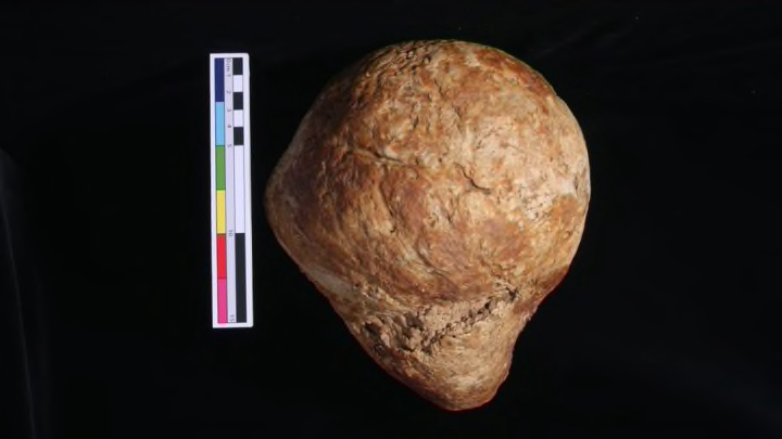
During recent excavations at a cemetery in southeast England, archaeologists pulled something strange out of an otherwise unremarkable grave. The object looked like a hybrid of a soccer ball and a rugby ball—bulbous at one end, tapered at the other. It was smooth as bone, resting near the hips of the skeleton of an older woman, who had been buried in a shroud at least 200 years ago.
“The first thing you would think is somehow the head has rolled down into the pelvis,” said Carolyn Rando, a forensic anthropologist at University College London. But the object wasn’t a skull. It was completely solid, and, at more than seven pounds, it was strikingly heavy. After making a careful analysis, Rando and her colleagues think it’s a calcified uterus, the largest of its kind in the archaeological record.
"I’ve never seen anything quite like that before, nor have my colleagues, and we were very excited,” Rando told mental_floss. “It’s one of the largest masses found archaeologically."
This giant calcified growth was found at St. Michael’s Litten, a graveyard in Chichester that was used from the Middle Ages until the mid-19th century, but had been hidden under a parking lot until excavations in 2011 turned up nearly 2000 bodies.
The uterus belonged to a woman who was over 50, had lost all of her teeth and had developed osteoporosis by the time she died, likely sometime between the 1600s and 1800s. (Archaeologists don’t have good dates for most of the graves at this cemetery.) The mass probably started out as a number of leiomyomas, sometimes called uterine fibroids, which are benign growths that occur in up to 40 percent of women of reproductive age. Most of the time, these masses remain soft tissue and don’t calcify. But some leiomyomas can get so large that they outstrip their blood supply and start to harden.
Photo courtesy of G. Cole, C. Rando, L. Sibun, and T. Waldron; UCL Institute of Archaeology
Rando and her colleagues came up with this diagnosis after conducting CT scans of the mass and then slicing it in half to look at its interior structure. In their case report, published in the September issue of the International Journal of Paleopathology, the scientists ruled out a long list of other potential conditions, including the possibility that the growth was a lithopedion, a fetus that dies during pregnancy and hardens outside the uterus. (This phenomenon occasionally shows up in the news, most recently in June, when a 50-year-old stone baby was found inside of an elderly woman in Chile.)
It’s not exactly clear how the growth affected the life of the woman who was buried at St. Michael’s, or if it contributed to her death.
“I’m sure she knew she had something,” Rando said. “I imagine that she might have had some problems going to the bathroom properly. I don’t think she would have been very comfortable. It would be like carrying a full-term infant all the time. But she lived a long life and this object would have taken a long time to grow, so maybe it didn’t bother her that much.”
In archaeological medical cases like this one, it’s tough to look for modern analogs, as most women today would get leiomyomas removed quite early, Rando said. But while scouring the historical medical literature, Rando and her colleagues did find one case that might shed light on how a woman could have lived with a baby-sized, calcified uterus for so long—and at what health risk. In 1840, a British doctor described a 72-year-old woman who came to him with intense abdominal pain after a fall. He noticed that she had a hard mass in her abdomen, which she said had been there for at least 30 years without causing her any trouble. Soon after the exam, the woman died. An autopsy revealed a tumor as hard as marble that resembled the uterus at five months pregnant, in both size and shape. The fall had caused this growth to perforate a section of the woman’s bowel, which killed her.
