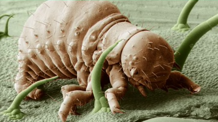
Thanks to electron microscopes, we can get an extremely close-up view of the wonder—or the horror—of the world around us. Here are a few images that just might fuel your nightmares.
1. Schistosome Parasite

Bruce Wetzel and Harry Schaefer via Wikimedia Commons
The schistosome parasite—magnified 256 times—lives in certain types of freshwater snails. It emerges into the water, where it can live for up to 48 hours. And now, for the horrifying part: The parasite can penetrate the skin of people who come into contact with contaminated water. After several weeks, the parasites mature into adult worms, which live and produce eggs in blood vessels.
Most people with schistosomiasis show no early signs, but "may develop a rash or itchy skin. Fever, chills, cough, and muscle aches can begin within 1-2 months of infection," according to the CDC. And that's just the beginning of the terrible havoc the eggs of this parasite wreak:
Eggs that are produced usually travel to the intestine, liver or bladder, causing inflammation or scarring. Children who are repeatedly infected can develop anemia, malnutrition, and learning difficulties. After years of infection, the parasite can also damage the liver, intestine, lungs, and bladder. Rarely, eggs are found in the brain or spinal cord and can cause seizures, paralysis, or spinal cord inflammation.
The parasites that cause schistosomiasis aren't found in the U.S., but 200 million people are infected worldwide.
2. Thaumetopoea processionea
This oak processionary caterpillar—which can be found in central and Southern Europe, and as far north as Sweden—has been magnified 30 times. In addition to posing a threat to oak trees, they also bother humans: Those little hairs (also called setae) are poisonous, causing asthma and skin irritation. According to a UK forestry website, they get their name because of their "distinctive habit of moving about in late spring and early summer in nose-to-tail processions."
3. Fruit Fly

Drosophila melanogaster—a common fruit fly, also called a vinegar fly—is often used in scientific research, and was one of the first organisms used for genetic analysis. The complete fruit fly genome was sequenced and published in 2000.
4. Cimex lectularius

Also known as a bed bug. This particular image, snapped by a scanning electron micrograph and digitally colorized, shows the insect's mouthparts, which it uses to pierce skin and drink your blood while you sleep. C. lectularius prefers to dine on human blood, but there are other types of bed bugs that dine exclusively on other animals, like poultry and bats.
Another fun fact about bed bugs: They mate using traumatic insemination. According to the paper "Reducing a cost of traumatic insemination: female bedbugs evolve a unique organ" published in a 2003 issue of the The Royal Society,
The male pierces the abdomen of the female with a sclerotized, needle-like paramere and inseminates into her body cavity despite the prescence of a fully functional female reproductive tract ... The females of C. lectularius (and most other cimicids) possess an organ called the spermalege. It has two embryologically discrete parts: the ectodermal ‘ectospermalege’ and the mesodermal ‘mesospermalege’. The ectospermalege consists of a groove in the right-hand posterior margin of the fifth sclerite overlying a structurally modified pleural membrane. During traumatic insemination, male bedbugs insert their intromittent organ into this groove, pierce the pleural membrane and so gain access to the female’s haemocoel (body cavity). ... Attached to the wall of the haemocoel, directly underneath the external groove, lies its second component: the mesospermalege. During traumatic insemination, sperm and seminal fluid are ejaculated into this haemocyte-containing membrane bound sac. Sperm travel out of the posterior part of the mesospermalege into the female’s haemolymph (blood) from where they migrate to specialized sperm storage structures (the seminal conceptacles) and then on to the ovaries where fertilization takes place.
So ... there you go.
5. Mosquito

This image, captured by a scanning electron microscope, shows an Anopheles gambiae mosquito magnified 114 times. A. gambiae is actually seven different species of mosquito that are indistinguishable from each other; this is called a complex.
6. Colorado Potato Beetle Nymph

Image courtesy of BARC/USDA
This image, snapped by a low temperature scanning electron microscope (LTSEM), shows a frozen first instar nymph of the Leptinotarsa decemlineata—also known as the Colorado Potato Beetle—magnified 100 times. As you might infer from its name, this bug loves potato crops and destroys plenty of them (and sometimes eggplant and tomato crops, too).
7. Head Louse
Looks kind of like a sloth, doesn't it? A sloth that climbs through your hair (and sometimes your eyebrows and eyelashes) laying eggs. Adult lice are just 2 to 3 mm long; this one has been magnified 200 times. The CDC estimates that, in the U.S., there are between 6 and 12 million cases of lice infestation in children 3 to 11 years old annually.
8. Yellow or Citrus Mite

Behold Lorryia formosa, the yellow mite. This head-on view of the citrus pest—which is falsely colored—was captured by freezing the mite and using a scanning electron microscope magnified 850 times. You can see a top view here.
9. Water Bear

ESA/Dr. Ralph O. Schill via NASA
Also known as Tardigrades, water bears are segmented, water-dwelling extremophiles (they're so extreme that even the vacuum of space is no big deal). When fully grown, they're just .5mm long. The critters were first described by German pastor J.A.E. Goeze in 1773; he called them kleiner Wasserbär, which means "little water bear." Perhaps Stephen Gammell used them as inspiration for his Scary Stories to Tell in the Dark illustrations?
10. Grub

Even in extreme close up, this grub will never be as horrifying as this giant variety, which turn into rhinoceros beetles.
11. Brown Recluse Spider

This pleasant looking guy, photographed at 27 times its normal size by scanning electron micrograph, was found in a Kentucky barn. Unlike other spiders—which have four pairs of eyes—Loxosceles reclusa has only three, and though tiny (typically under an inch), it packs serious bite: Brown recluses have potentially deadly hemotoxic venom, which can sometimes cause necrosis of the skin.
BONUS: Snail Love Dart

Sure, "love dart" sounds romantic, but hermaphrodite snails use these parts to stab each other while mating. Not so sweet now, huh? This particular dart comes from the white-lipped snail (Cepaea hortensis) and looks like something straight out of a horror movie.
