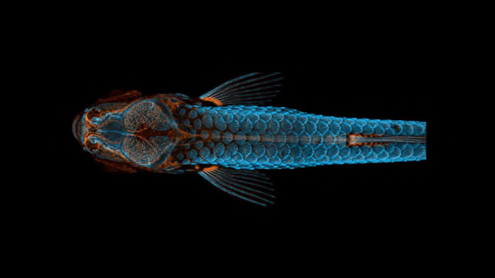
Each year, Nikon holds the Nikon Small World Photomicrography Competition to recognize the coolest images of the smallest subjects. The 2020 winners were just announced, and as usual, it’s a stunning collection of things only visible under a microscope—hippocampal neurons, for example—and familiar things that look completely different under a microscope (like human hair).
First place went to Daniel Castranova, an aquatic research specialist at the National Institute of Health, for his snapshot of a juvenile zebrafish. The photo isn’t the result of a brief camera click; instead, Castranova and his colleague Bakary Samasa scanned the fish using a technique called “confocal microscopy” and then stacked more than 350 frames to create one comprehensive image. During the project, the researchers realized that zebrafish—which are already used as lab models to study many human diseases—have lymphatic vessels in their skulls, which means their lymphatic systems are much more similar to humans’ than previously thought. They're so similar that zebrafish may prove useful in Alzheimer’s disease and cancer research.
“Until now, we thought this type of lymphatic system associated with the nervous system only occurred in mammals,” Castranova told Nikon. “By studying them, the scientific community can expedite a range of research and clinical innovations—everything from drug trials to cancer treatments. This is because fish are so much easier to raise and image than mammals.”
The image is also evidence that art and science can go hand in hand—the overarching point of the whole competition. See some of our other favorite winners below, and scroll through the full gallery here.
Embryonic Development of a Clownfish // Second Place

Daniel Knop from Germany’s Natur und Tier Verlag stacked images to capture the progression of a clownfish (Amphiprion percula) forming in its egg—a difficult feat, considering that the embryo didn’t exactly stop moving to pose.
Tongue of a Freshwater Snail // Third Place

A snail’s tongue, or radula, is covered in thousands of microscopic teeth that rub against its food to snag off small bits. As demonstrated by this photo from Howard Hughes Medical Institute’s Dr. Igor Siwanowicz, it’s actually more dazzling than disgusting.
Bogong Moth // Fifth Place

The dull brown coat of Australia’s Bogong moth is nothing special from afar. Magnified under the lens of Indonesia-based microphotographer Ahmad Fauzan, it looks like a tiger-inspired shag carpet (which is almost as cool as its spiral tongue).
Red Algae // 11th Place

Red algae’s tendrils have a skeletal quality even when viewed with the naked eye. University of Maryland, Baltimore County’s Dr. Tagide deCarvalho shows just how much they look like long, alien skeleton hands when seen beneath a microscope.
Crystals Formed From an Ethanol and Water Solution // 13th Place

New York-based photographer Justin Zoll used polarized light to reveal the vibrant details of crystals created when an amino acid-containing solution of ethanol and water was heated.
Nylon Stockings // 16th Place

Micrography can also illuminate the beauty of seemingly mundane, human-made products. This image, taken by Alexander Klepnev at Moscow’s JSC Radiophysics, shows how nylon fibers are knotted to make a pair of tights.
Skeleton of a Fruit Bat Embryo // 20th Place

University of Cape Town’s Dr. Dorit Hockman and Dr. Vanessa Chong-Morrison didn’t utilize any light-filtering techniques to snap this photo of a short-tailed fruit bat's embryonic skeleton smiling at you (or so it seems). Happy Halloween!
